Eye Care Technology
Good equipment doesn’t replace a good doctor, but it does let one see more. Here’s what we use, and what each piece is actually for.
Digital retinal photography
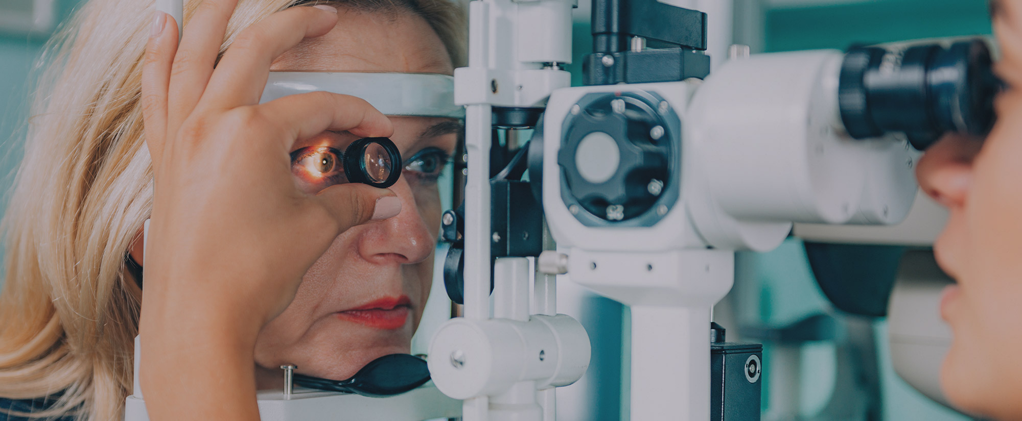
Retinal photography uses a specialized low-power microscope with an attached camera to photograph the interior surface of the eye: the retina, the optic disc and the macula. It lets us diagnose eye disease and then track whether it’s progressing. The conditions we most often find and monitor this way are glaucoma, macular degeneration, diabetic retinopathy and hypertensive retinopathy.
We also take a baseline image of your retina to compare against in future years, which is one of the most useful things preventive eye care can do. Our retinal camera is networked into every exam room, so we can put the photo on a screen and show you what we’re talking about rather than describing it.
Topcon TRC-NW400 fundus camera
This is the camera behind those images. It’s a non-mydriatic fundus camera, meaning it produces true-color photos of the retina in high quality without requiring dilation first. Detail is clear and sharp. It has the traditional three fixation targets for nasal, central and temporal images, plus nine more for peripheral imaging, so we can capture the full periphery as well. That lets us do either a quick screening or a more thorough examination on the same instrument. Fundus cameras of this type are approved for diabetic eye screening.
Glaucoma pressure testing
Topcon CT-80 computerized non-contact tonometer
This is the “puff of air” test, and this model makes it easier and more comfortable than it used to be. Measurement is about 30% faster, with a triple patient safety function and a dual sensor system for detecting and monitoring glaucoma. If you’re a glaucoma patient or a glaucoma suspect, we’ll also do applanation tonometry for a more precise reading of intraocular pressure.
Visual field testing
Humphrey FDT: frequency doubling technology
A visual field test checks your peripheral, or side, vision. Field loss can come from disease affecting the eye, the optic nerve or the brain. The test flashes points of light onto a white screen and you press a button whenever you see one; the computer maps and calculates your field from your responses.
Our doctors use visual fields to diagnose and monitor glaucoma, to pick up central or peripheral retinal disease, to assess eyelid conditions such as ptosis, and to check for optic nerve disease, problems along the visual pathways in the brain, tumors, and retinal toxicity from hydroxychloroquine (Plaquenil).
Our own lens lab
Optronics lens edger
We cut lenses on site. The Optronics edger handles all shapes, sizes and powers of CR-39, polycarbonate and hi-index lenses, and we stock digital, plastic, polycarbonate, high index, progressive and flat-top bifocal lenses.
The practical upshot: same-day service on most single vision lenses, and next-day service on most progressives and bifocals. Most places send that work out and you wait a week.
Digital documentation
Topcon DC-3 digital camera
Eight megapixels, for clear images and video and high-quality digital records of what we find.
Your records, online
We run RevolutionEHR for medical billing, scheduling, image archiving and accounting. It also gives you a Personal Health Record, so you can look up your own appointments, billing, prescriptions and other information in your file rather than calling to ask.
Book an exam
All of the above is standard equipment in a comprehensive exam here, not an upsell. Request a time online, or call (843) 796-1155.
Request an appointment · What an eye exam covers · Eye conditions we treat